
Young researcher has created a sensor that detects errors in MRI scans
2.5.2024 12:33:55 CEST | Københavns Universitet | Press release
Hvidovre Hospital has the world's first prototype of a sensor capable of detecting errors in MRI scans using laser light and gas. The new sensor, developed by a young researcher at the University of Copenhagen and Hvidovre Hospital, can thereby do what is impossible for current electrical sensors – and hopefully pave the way for MRI scans that are better, cheaper and faster.
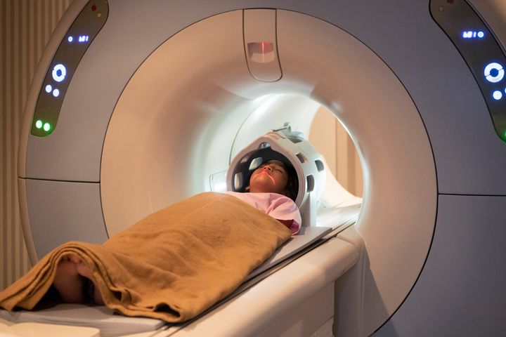
MRI scanners are used by doctors and healthcare professionals every day to get a unique look into the human body. In particular, they are used to study the brain, vital organs and other soft tissues by way of 3D images of exceptional quality compared to other types of medical imaging.
While this makes the advanced tool invaluable and nearly indispensable for healthcare professionals, there is still room for improvement.
The strong magnetic fields inside MRI scanners have fluctuations that create errors and disturbances in scans. Consequently, these expensive machines (hundreds of Euros per hour) must be calibrated regularly to reduce errors.
There are also special scanning methods, which unfortunately cannot be done in practice today. Among them, so-called spiral sequences that could reduce scanning time, e.g., when diagnosing blood clots, sclerosis and tumors. Spiral sequences would also be an attractive tool in MRI research, where, among other things, they could provide researchers and health professionals with new knowledge about brain diseases. But due to the highly unstable magnetic field, performing these types of scans is not currently an option.
In theory, the problem can be solved with a sensor that reads and maps changes in the magnetic field. Thereafter, it is relatively simple to correct the errors in images with a computer. In practice, this has been difficult with the current technology, as otherwise suitable sensors interfere with the magnetic field because they are electric and connected to metal cables.
A new invention hopes to make this problem a thing of the past. To combat the problem, a researcher from the Niels Bohr Institute and The Danish Research Centre for Magnetic Resonance (DRCMR) has developed a sensor that uses laser light in fiber cables and a small glass container filled with gas. The prototype is ready and works.
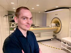
"First we demonstrated that it was theoretically possible, and now we have proven that it can be done in practice. In fact, we now have a prototype that can basically make the measurements needed without disturbing the MRI scanner. It needs to be developed more and fine-tuned, but has the potential to make MRI scans cheaper, better and faster – although not necessarily all three at once," laughs Hans Stærkind, a postdoc at the Niels Bohr Institute and DRCMR at Hvidovre Hospital. Stærkind is the main architect behind the sensor and device that comes with it.
"An MRI scanner can already produce incredible images if one takes their time. But with the help of my sensor, it is imaginable to use the same amount of time to produce even better imagery – or spend less time and still get the same quality as today. A third scenario could be to build a cheaper scanner that, despite a few errors, could still deliver decent image quality with the help of my sensor," says the researcher.
How the prototype works
MRI scanners use powerful magnets to produce a strong magnetic field that forces protons in the body's water, carbohydrates and proteins to align themselves with the magnetic field. When radio waves are pulsed through a patient, the protons are stimulated and temporarily spin out of that equilibrium. When they subsequently return to alignment with the magnetic field, they release radio waves that can be used to form real-time 3D images of whatever is being scanned.
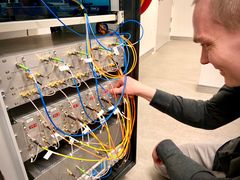
Hans Stærkind's prototype works using a device for sending and receiving laser light that looks like a 1990’s stereo system. It sends laser light through fiber optic cables – i.e., without any metal – and into four sensors located in the scanner.
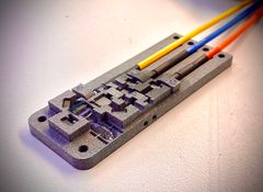
Within the sensors, the light passes through a small glass container containing a caesium gas, which absorbs the light at the right light frequencies.
"When the laser has just the right frequency while passing through the gas, there is a resonance between the waves of light and electrons in the caesium atoms. But the frequency – or wavelength – at which this happens changes when the gas is exposed to a magnetic field. In this way, we can measure the strength of the magnetic field by finding out what the right frequency is. This happens completely automatically and lightning fast by the receiving device," explains the researcher.
As disturbances in an MRI scanner's ultra-powerful magnetic field occur, Hans Stærkind's prototype maps where in the magnetic field they are occurring and by what strength the field has changed. In the near future, this could mean that disturbed and faulty images could be corrected – based on the data collected by the sensors, and subsequently made accurate and entirely usable.
Innovation with commercial prospects – when data is in place
The prototype is currently housed at DRCMR at Hvidovre Hospital in Copenhagen, which is also where the idea was conceived.
"The original idea came from my supervisor here at DRCMR, Esben Petersen, who is unfortunately no longer with us. He saw huge potential in developing a sensor based on lasers and gas that would be able to measure the magnetic fields without disturbing them," says Hans Stærkind.
With the help of quantum physicists at the Niels Bohr Institute, including Professor Eugene Polzik, Stærkind developed the idea into an actual theory. And with the prototype, he has now put that theory into practice.
"The prototype is designed in such a way that it is already suitable in hospital contexts as a robust and well-functioning instrument. And so far, our tests have shown that it works as it should. One can imagine that this invention will eventually be integrated directly into new MRI scanners," says Stærkind.
For now, the prototype will be developed further so that its measurements become even more accurate.
"We need to collect data and fine-tune it so that it continuously becomes a better and better tool for finding errors in scans. After that, we’ll move on to the exciting work of correcting errors in MRI images, and find out in what situations and which types of scans our sensor can make a significant difference," says the researcher.
According to Stærkind, the immediate target group for his sensor are MRI research units. But he also hopes that one of the large MRI manufacturers finds out about the new technology, in the slightly longer term.
"Once the prototype has been refined in a 2.0 version and its qualities documented with plenty of data from actual scans here at the hospital, we will see where this goes. It certainly has the potential to improve MRI scans in a unique way that can benefit doctors and, not least, patients," says the researcher.
*
Extra info: Facts about MRI scanners
Despite having been around since 1977, MRI scanners remain one of the most advanced medical technologies. In fact, everything from quantum mechanics, superconducting magnets and advanced mathematics and computer science is a prerequisite for them to work.
The devices consist of a giant magnet with a magnetic force so great, that it must be cooled to -269° C or risk going up in smoke – literally. Among other things, this is done with liquid helium and makes the machine's primary magnet superconductive.
That is, the electricity that drives electromagnetism has no resistance, and constantly runs in a closed circuit without the supply of electricity. The whopping electric bills associated with operating MRI’s are primarily due to their cooling.
Within an MRI scanner, there are a number of other electromagnets that can be used to control the magnetic field, so that you can look into specific parts of the body and do so from different angles.
The very high strength of the magnetic fields requires that belt buckles, coins and all other metal objects be kept safely away from the machine in another room. In fact, a number of accidents with MRI scanners have occurred due to their exceptionally powerful magnetism. For example, a wheelchair could be hurled towards the scanner regardless of who or what was standing in its way. But if all of the necessary safety precautions are followed, there are no known risks from an MRI scan itself.
The scanner's strong magnetic field forces protons in the body's water molecules – which are themselves magnets, called spins – to align themselves with the magnetic field. Radio waves are then sent through the patient, which temporarily spin the protons out of that equilibrium. When realigned, the energy is released again in measurable radio waves.
With the help of a computer, magnetic resonance imaging (MRI) can be used to create millimetre-precise 3D images of a patient’s soft tissue from any angle.
Facts: How it works
Four sensors are distributed in the MRI scanner. One remains out of range of the magnetic field and acts as a control.
Laser light inside the sensors with certain light frequencies passes through a small glass container with cesium gas.
The frequency of the laser creates resonance in the electrons of the cesium atoms. This dims the light to a degree that can be detected.
If the gas is exposed to a magnetic field, the triggering frequency changes depending on the strength of the magnetic field.
Fluctuations in the magnetic field of the MRI scanner can thus be registered and data can subsequently reveal errors in the MRI scan.
Facts: Resonance
In the Adventures of Tintin, opera diva Bianca Castafiore shatters a crystal glass by hitting the glass’s resonant frequency with the power of her voice. Everything has a certain frequency that it likes to vibrate – or oscillate at.
If as a child, or adult, you ever set a swing in motion by pumping it back and forth, you used the resonance frequency to do so. When something resonates, its oscillations are amplified.
If you send light into a gas, it will pass straight through – unless it has just the right frequency. At a certain frequency, light is absorbed because it oscillates at the same frequency as the electrons in the gas atoms.
The electrons oscillate more and more while absorbing the energy, and the light is then re-emitted in all directions as the electrons fall back into place.
If you look at it, you will see that the ray dims and the gas vapor lights up.
Resonance, therefore, is when you hit the natural frequency of a system so that it oscillates. This frequency is called resonance frequency.
About the project: part of a new research center
The sensor will continue development at Copenhagen Center for Biomedical Quantum Sensing, a research center funded by the Novo Nordisk Foundation and headed by Professor Eugene Polzik.
The prototype is being tested in collaboration with the Danish Research Centre for Magnetic Resonance, at Hvidovre Hospital
Behind the research:
From the Niels Bohr Institute, University of Copenhagen:
Hans Stærkind
Kasper Jensen
Jörg H. Müller
and Eugene S. Polzik
From the The Danish Research Centre for Magnetic Resonance (DRCMR) at Hvidovre Hospital:
Vincent O. Boer
and Esben T. Petersen
Keywords
Contacts
Hans StærkindPostdocNiels Bohr Institute, University of Copenhagen
Tel:+45 50574438hans.staerkind@nbi.ku.dkKristian Bjørn-HansenJournalist and Press ContactFaculty of Science, Copenhagen University
Tel:+45 93516002kbh@science.ku.dkImages

Links
ABOUT THE FACULTY OF SCIENCE
The Faculty of Science at the University of Copenhagen – or SCIENCE – is Denmark's largest science research and education institution.
The Faculty's most important task is to contribute to solving the major challenges facing the rapidly changing world with increased pressure on, among other things, natural resources and significant climate change, both nationally and globally.
Subscribe to releases from Københavns Universitet
Subscribe to all the latest releases from Københavns Universitet by registering your e-mail address below. You can unsubscribe at any time.
Latest releases from Københavns Universitet
Fortidens massive hvalfangst truer fremtidens grønlandshvaler17.3.2026 16:00:00 CET | Pressemeddelelse
Kommerciel hvalfangst har gjort grønlandshvalen sårbar 1.000 år ud i fremtiden.
AI skulle lette lægernes arbejde – i stedet bruger de timer på at rette fejl17.3.2026 12:44:38 CET | Pressemeddelelse
AI-baseret journalisering skulle effektivisere arbejdet på landets hospitaler. Men i praksis ender læger med at bruge værdifuld arbejdstid på at rette fejl, træne algoritmer og løse administrative opgaver, som før lå hos sekretærerne, viser ny forskning fra Københavns Universitet.
Mørke personlighedstræk har betydning for valg af karriere10.3.2026 05:45:38 CET | Pressemeddelelse
Personer med høj andel af såkaldte mørke personlighedstræk har betydeligt mindre interesse for sociale og kreative jobs end andre mennesker har. Det viser ny forskning fra Københavns Universitet.
Københavns Universitet lancerer stor vidensportal til kvinder i overgangsalderen5.3.2026 11:00:00 CET | Pressemeddelelse
Mange kvinder mangler viden om overgangsalderen, som længe har været et underprioriteret emne. En pjece og en ny vidensportal fra forskningsinitiativet Kvinder i Sund Overgang (KiSO) ved Københavns Universitet skal hjælpe kvinder med at få overblik og forstå denne fase af livet.
Her er Københavns Universitets valgeksperter4.3.2026 16:14:11 CET | Pressemeddelelse
Folketingsvalget er i gang. Der er stor efterspørgsel på ekspertkilderne. Her er Københavns Universitets liste med et udpluk af forskere, der kan agere eksperter i valgkampen.
In our pressroom you can read all our latest releases, find our press contacts, images, documents and other relevant information about us.
Visit our pressroom